


Summary: Researchers created the largest 3D reconstruction of human brain tissue at synaptic resolution, capturing detailed images of a cubic millimeter of human temporal cortex. This tiny piece of brain contains 57,000 cells, 230 millimeters of blood vessels, and 150 million synapses, which amounts to 1,400 terabytes of data.
This research is part of a broader effort to map an entire mouse brain’s neural wiring, with hopes of advancing our understanding of brain function and disease. The technology combines high-resolution electron microscopy and AI-powered algorithms to meticulously color-code and map out the complex neural connections.
Key Facts:
> Intricate Details: The reconstructed brain tissue includes unprecedented details, such as a unique set of axons connected by up to 50 synapses, and other peculiar formations potentially linked to epilepsy in the patient from whom the sample was taken.
Massive Data Volume: The study tackled a piece of brain tissue containing a data volume equivalent to 1,400 terabytes, illustrating the complexity and data-intensive nature of mapping neural connections.
Technological Integration: The research utilized cutting-edge AI algorithms developed by Google to assist in the 3D reconstruction of the brain tissue, highlighting a significant collaboration between biological research and technological innovation.
Source: Harvard
A cubic millimeter of brain tissue may not sound like much. But considering that tiny square contains 57,000 cells, 230 millimeters of blood vessels, and 150 million synapses, all amounting to 1,400 terabytes of data, Harvard and Google researchers have just accomplished something enormous.
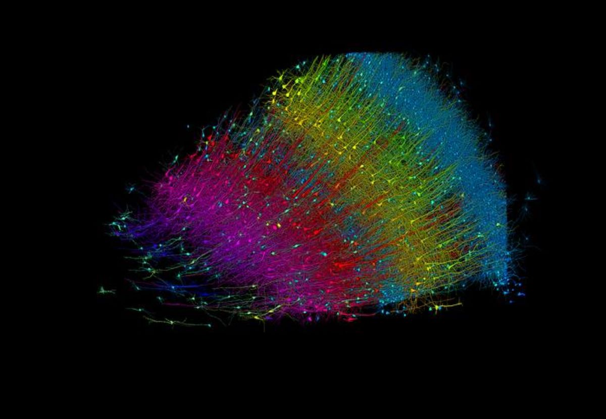
A Harvard team led by Jeff Lichtman, the Jeremy R. Knowles Professor of Molecular and Cellular Biology and newly appointed dean of science, has co-created with Google researchers the largest synaptic-resolution, 3D reconstruction of a piece of human brain to date, showing in vivid detail each cell and its web of neural connections in a piece of human temporal cortex about half the size of a rice grain. Six layers of excitatory neurons color-coded by depth. Credit: Google Research and Lichtman Lab The feat, published in Science, is the latest in a nearly 10-year collaboration with scientists at Google Research, who combine Lichtman’s electron microscopy imaging with […]

Get Creative with Cannabis: The Ultimate Guide to Painting and Pottery Workshops
Creative with Cannabis Cannabis and creativity go hand in hand—especially


